


.png)

 ( 母液浓度为 40 mg/mL ), 如您需要配置的浓度超过该产品的溶解度,请先与我们客服联系。
| 中文名称: | CHR-6494 一键复制产品信息 | ||||
|---|---|---|---|---|---|
| 英文名称: | CHR-6494 | ||||
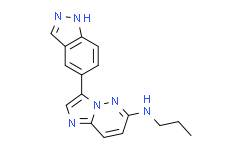
| 别名: | CHR-6494 国华试剂;3-(1H-吲唑-5-基)-N-丙基咪唑并[1,2-b]哒嗪-6-胺;3-(1H-吲唑-5-基)-N-丙基咪唑并[1,2-B]哒嗪-6-胺;3-(1H-吲唑-5-基)-n-丙基-咪唑并[1,2-b]吡嗪-6-胺 CHR 6494;CHR-6494 trifluoroacetate salt;3-?(1H-?indazol-?5-?yl)?-?N-?propyl-Imidazo[1,?2-?b]?pyridazin-?6-?amine;3-(1H-Indazol-5-yl)-N-propylimidazo-[1,2-b]pyridazin-6-amine;CHR-6494 | ||||
| CAS No: | 1333377-65-3 | 分子式: | C16H16N6 | 分子量: | 292.34 |
| CAS No: | 1333377-65-3 | ||||
| 分子式: | C16H16N6 | ||||
| 分子量: | 292.34 | ||||
包装规格:
5mg 10mg 25mg 50mg 100mg in glass bottle
溶解性:
溶于DMSO(50mg/mL超声)
产品描述:
基本信息
|
产品编号: |
C10925 |
||||
|
产品名称: |
CHR-6494 |
||||
|
CAS: |
1333377-65-3 |
储存条件 |
粉末 |
-20℃ |
四年 |
|
|
|
||||
|
分子式: |
溶于液体 |
-80℃ |
六个月 |
||
|
分子量 |
292.34 |
-20℃ |
一个月 |
||
|
化学名: |
3-(1H-Indazol-5-yl)-N-propylimidazo-[1,2-b]pyridazin-6-amine;CHR-6494 |
||||
|
Solubility (25°C): |
|||||
|
体外:
|
DMSO |
50mg/mL(171.03mM;Need ultrasonic) |
|||
|
Ethanol |
|
||||
|
Water |
|
||||
|
体内(现配现用): |
1.请依序添加每种溶剂:10% DMSO→40% PEG300→5% Tween-80→45% saline Solubility:≥2.5mg/mL(8.55mM);Clear solution |
||||
|
此⽅案可获得≥2.5mg/mL(8.55mM,饱和度未知)的澄清溶液。以1mL⼯作液为例,取100μL25.0mg/mL的澄清DMSO储备液加到400μL PEG300中,混合均匀;向上述体系中加⼊50μL Tween-80,混合均匀;然后继续加⼊450μL⽣理盐⽔定容⾄1mL。 |
|||||
|
2.请依序添加每种溶剂:10% DMSO→90% corn oil Solubility:1mg/mL(3.42mM);Suspended solution; Need ultrasonic |
|||||
|
此⽅案可获得1mg/mL(3.42mM)的均匀悬浊液,悬浊液可⽤于⼝服和腹腔注射。以1mL⼯作液为例,取100μL10.0mg/mL的澄清DMSO储备液加到900μL⽟⽶油中,混合均匀。 |
|||||
|
<1mg/ml表示微溶或不溶。 |
|||||
|
普西唐提供的所有化合物浓度为内部测试所得,实际溶液度可能与公布值有所偏差,属于正常的批间细微差异现象。 |
|||||
|
请根据产品在不同溶剂中的溶解度选择合适的溶剂配制储备液;⼀旦配成溶液,请分装保存,避免反复冻融造成的产品失效。 |
|||||
制备储备液
|
浓度
溶液体积 质量 |
1mg |
5mg |
10mg |
|
1mM |
3.4207mL |
17.1034mL |
34.2067mL |
|
5mM |
0.6841mL |
3.4207mL |
6.8413mL |
|
10mM |
0.3421mL |
1.7103mL |
3.4207mL |
生物活性
|
产品描述 |
一种有效的 haspin 抑制剂,能够抑制组蛋白 H3T3 的磷酸化,IC50 值为 2nM。 |
|
靶点 |
haspin 2nM (IC50) |
|
体外研究 |
CHR-6494 (0-10-5nM; 72 hours) dose-dependently inhibits the growth of cancer cells, such as HCT-116, HeLa, MDA-MB-231, and Wi-38 cells, with IC50s of 500nM, 473nM, 752nM and 1059nM, respectively. CHR-6494 (500nM) produces a mitotic catastrophe with abnormal morphology of the mitotic spindle and centrosome amplification, and upregulates the spindle assembly checkpoint protein BUB1 and the marker of mitotic arrest cyclin B1. CHR-6494 exhibits inhibitory activities against melanoma cell lines, including BRAFV600E mutants, NRAS mutants, and wild type cells, with IC50s ranging from 396nM to 1229nM. CHR-6494 (300nM and 600nM; 72 hours) induces apoptosis, increases caspase 3/7 activity by 3- and 6-fold, respectively in COLO-792 cells, and to 8.5- and 16-fold in RPMI-7951 cells. CHR-6494 in combination with MEK inhibitors synergistically inhibits viability of melanoma cells, enhances apoptosis in melanoma cells, modulates cell cycle progression independently by arresting melanoma cells at different phases, and suppresses migration of melanoma cells. CHR-6494 (50, 200nM; 1 week) enhances the antiproliferative effects of MLN8237 in MDA-MB-231, SKBR3 breast cancer cells. CHR-6494 (200 nM; 72 hours) enhances the apoptosis of MDA-MB-231 and SKBR3 cells when combined with MLN8237 |
|
体内研究 |
CHR-6494 (50mg/kg; i.p. in two cycles of five consecutive days for 15 days) inhibits the growth of tumor and cuases no obvious body weight change in nude mice bearing HCT-116 human colorectal cancer cells. CHR-6494 (20mg/kg; intraperitoneal injection for 15 consecutive days) inhibits the tumor volume and weight compared with the control group in nude mice bearing MDA-MB-231 xenograft tumors. CHR-6494 (20mg/kg; intraperitoneal injection for 15 consecutive days) enhances the tumor volume and weight inhibition of MLN8237 (20 mg/kg; p.o.) in vivo |
推荐实验方法(仅供参考)
|
Kinase Assay |
The analysis of the enzymatic inhibitory capacity of the compound in a panel of 29 protein kinases is developed using a FRET assay based on the differential sensitivity of phosphorylated and non-phosphorylated peptides to protein cleavage (Z′-LYTE Kinase Assay). In the primary reaction, the kinase transfers the γ-phosphate of ATP to a single tyrosine, serine or threonine residue in a synthetic FRET peptide. In the secondary reaction, a site-specific protease recognizes and cleaves non-phosphorylated FRET peptides. Phosphorylation of FRET peptides suppresses cleavage by the development reagent. Cleavage disrupts FRET between the donor (coumarin) and the acceptor (fluorescein) fluorophores on the FRET peptide, whereas uncleaved, phosphorylated FRET peptides maintain FRET. A ratiometric method, which calculates the ratio (the emission ratio) of donor emission to acceptor emission after excitation of the donor fluorophore at 400n, is used to quantitate reaction progress |
|
|
|
|
Cell Assay |
Cells are treated for 24, 48 and 72 h with the inhibitor or with DMSO as a control. Cell viability is assessed using the colorimetric XTT assay. Cells are seeded in 94-well plates at a density of 4 × 10⁴ cells per well, and allowed to attach for 24h.The medium is then exchanged with others containing different drug concentrations (0.001−10μM). Eight wells for each concentration of the CHR-6494 compound are used. At the corresponding time, the culture medium is discharged, the XTT reagent is added and the final cell number and optical density are determined. Dose-response curves are generated and cell viability is evaluated after 72h of treatment. The half-maximal inhibitory concentration (IC50) is determined using GraphPad Prism software |
|
|
|
|
Animal Administration |
Athymic nu/nu male mice, aged 4-5 weeks, are used for tumor xenograft assays. Animals are maintained in a sterile environment; their cages, food and bedding are sterilized by autoclaving. Mice are anesthetized and tumor cells are injected subcutaneously. In all, 3.5 × 10⁶ exponentially growing HCT-116 cells diluted in 250μL of sterile PBS are injected subcutaneously in each animal (n = 30). Body weight is recorded and tumor dimensions are measured twice weekly using digital calipers. Tumor volume (in mm3 ) is estimated according to the formula V = D × d2 /2, where D is the long axis and d the short axis of tumor. When tumors reach an average volume of 200 mm3 (15 days after injection), 24 mice harboring homogeneous tumor sizes are randomized into two groups: (1) control group (n = 8) treated with vehicle (solution of 10% DMSO/20% 2-hydroxypropyl-b-cyclodextrin; (2) treatment group (n = 16) mice is diary treated by intraperitoneal injection of 50 mg/kg of CHR-6494 diluted in a solution of 10% DMSO/20% 2-hydroxypropyl-b-cyclodextrin in two cycles of five consecutive days for 15 days. The treatment group is randomly divided into a short-time response group (n = 8), defined by tumor weight at the moment of killing of the control group, and a long-time response group (n = 8), defined by tumor regrowth after treatment. Mice are killed at the end of treatment, and tumors from both groups are excised and weighted. The mean volume of tumor mass is expressed as mean ± s.e.m. for each mouse group, and significance is assessed by means of the Mann-Whitney U-test. Values of P < 0.05 are considered statistically significant. Upon killing mice, colon, lung, liver and kidney tissues are obtained to analyze endogenous toxicity by hematoxylin and eosin |
保存条件:
-20℃
UN码:
HazardClass:
危害声明:
安全说明:
搜索质检报告(COA)
本计算器可帮助您计算出特定溶液中溶质的质量、溶液浓度和体积之间的关系,公式为:
质量 (g) = 浓度 (mol/L) x 体积 (L) x 分子量 (g/mol)
摩尔浓度计算公式
用本工具协助配置特定浓度的溶液,使用的计算公式为:
开始浓度 x 开始体积 = 最终浓度 x 最终体积
稀释公式
稀释公式一般简略地表示为:C1V1 = C2V2 ( 输入 输出 )
(2).jpg)
体内配方的制备方法: 取 50μLDMSO 母液,添加 300 μLPEG300 混匀澄清,再加 50μLTween80, 混匀澄清,再加 600μLSaline/PBS/ddH2O 混匀澄清
方案所需的各种助溶剂如: DMSO , PEG300 / PEG400 , Tween 80 , SBE-β-CD , 玉米油 等, 均可点击跳转或在网站搜索购买。
母液,添加 300 μLPEG300 混匀澄清,再加 50μLTween80, 混匀澄清,再加 600μLSaline/PBS/ddH2O 混匀澄清方案所需的各种助溶剂如: DMSO , PEG300 / PEG400 , Tween 80 , SBE-β-CD , 玉米油 等, 均可点击跳转或在网站搜索购买。
以上为“动物实验计算换算器”的使用方法举例,并不是具体某个试剂的配制,请根据您的实验动物和给药方式选择适当的溶解方案。
计算结果:
工作液浓度: mg/ml;
DMSO母液配制方法: mg 药物溶于 μL DMSO溶液(母液浓度 mg/mL,
体内配方配制方法:取 μL DMSO母液,加入 μL PEG300,混匀澄清后加入μL Tween 80,混匀澄清后加入 μL Saline/PBS/ddH2O,混匀澄清。
1. 首先保证母液是澄清的;
2.
一定要按照顺序依次将溶剂加入,进行下一步操作之前必须保证上一步操作得到的是澄清的溶液,可采用涡旋、超声或水浴加热等物理方法助溶。
剂量转换
对于不同动物的给药剂量换算,您也可以参考 更多


