
| 中文名称: | Ripasudil free base | ||||
|---|---|---|---|---|---|
| 英文名称: | Ripasudil free base | ||||
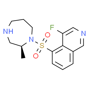
| 别名: | 4-fluoro-5-[[(2S)-2-methyl-1,4-diazepan-1-yl]sulfonyl]isoquinoline K-115 (free base) | ||||
| CAS No: | 223645-67-8 | 分子式: | C15H18FN3O2S | 分子量: | 323.39 |
| CAS No: | 223645-67-8 | ||||
| 分子式: | C15H18FN3O2S | ||||
| 分子量: | 323.39 | ||||
![2,4-二甲基苯甲酸 [4-[(1S)-1-(氨基甲基)-2-(6-异喹啉基氨基)-2-氧代乙基]苯基]甲基酯二盐酸盐](http:///www.jm-bio.com/content/xtheme/beijingjinming/images/noimage.jpg)
基本信息
|
产品编号: |
R10278 |
||||
|
产品名称: |
Ripasudil free base |
||||
|
CAS: |
223645-67-8 |
储存条件 |
粉末 |
-20℃ |
四年 |
|
|
|
||||
|
分子式: |
溶于液体 |
|
|
||
|
分子量 |
323.39 |
|
|
||
|
化学名: |
4-fluoro-5-[[(2S)-2-methyl-1,4-diazepan-1-yl]sulfonyl]isoquinoline K-115 (free base) |
||||
|
Solubility (25°C): |
|||||
|
体外:
|
DMSO |
|
|||
|
Ethanol |
|
||||
|
Water |
|
||||
|
体内(现配现用): |
|
||||
|
|
|||||
|
<1mg/ml表示微溶或不溶。 |
|||||
|
普西唐提供的所有化合物浓度为内部测试所得,实际溶液度可能与公布值有所偏差,属于正常的批间细微差异现象。 |
|||||
|
请根据产品在不同溶剂中的溶解度选择合适的溶剂配制储备液;⼀旦配成溶液,请分装保存,避免反复冻融造成的产品失效。 |
|||||
生物活性
|
产品描述 |
一种ROCK 特异性抑制剂,能够抑制 ROCK2 和 ROCK1 的活性,IC50 值分别为 19 和 51 nM。 |
||||
|
靶点 |
ROCK2 |
ROCK1 |
CaMKIIa |
PKACa |
PKC |
|
19nM (IC50) |
51nM (IC50) |
370nM (IC50) |
2.1μM (IC50) |
27μM (IC50) |
|
|
体外研究 |
Ripasudil (K-115) is a potent inhibitor of ROCK, with IC50s of 19 and 51 nM for ROCK2 and ROCK1, respectively. Ripasudil also shows less potent inhibitory activities against CaMKIIα, PKACα and PKC, with IC50s of 370 nM, 2.1μM and 27μM, respectively . Ripasudil (K-115; 1, 10μM) induces cytoskeletal changes, including retraction and cell rounding and reduced actin bundles of cultured trabecular meshwork (TM) cells. Ripasudil (5μM) sifnificantly reduces transendothelial electrical resistance (TEER), and increases FITC-dextran permeability in Schlemm’s canal endothelial (SCE) cell monolayers |
||||
|
体内研究 |
Ripasudil (K-115) reduces intraocular pressure (IOP) in a concentration-dependent manner at concentrations between 0.1% and 0.4% in monkey eyes and 0.0625% to 0.5% in rabbit eyes, respectively[1]. Ripasudil (K-115; 1mg/kg, p.o. daily) shows a neuroprotective effect on retinal ganglion cells (RGCs) after nerve crush (NC). Ripasudil also inhibits the oxidative stress induced by axonal injury in mice. Ripasudil suppresses the time-dependent production of ROS in RGCs after NC injury |
||||
推荐实验方法(仅供参考)
|
Kinase Assay |
ROCK 1 (0.75ng/mL) and ROCK 2 (0.5ng/mL) are incubated with various concentrations of Ripasudil, Y-27632, or HA-1077 at 25°C for 90 min in 50mM Tris-HCl buffer (pH 7.5) containing 100mM KCl, 10mMmgCl2, 0.1mM EGTA, 30mM Long S6 Kinase Substrate peptide, and 1mM ATP in a total volume of 40mL. PKACa, PKC, and CaMKIIa are also incubated with various concentrations of Ripasudil, Y-27632, or HA-1077. PKACa (0.0625ng/mL) is incubated at 25°C for 30 min in 40mM Tris-HCl buffer (pH 7.5) containing 20mMmgCl2, 1mg/mL BSA, 5mM Kemptide peptide substrate, and 1mM ATP in a total volume of 40mL. PKC (0.025ng/mL) is incubated at 25°C for 80 min in 20mM Tris-HCl buffer (pH 7.5) containing 20mMmgCl2, 0.4mM CaCl2, 0.1mg/mL BSA, 0.25mM EGTA, 25ng/mL phosphatidylserine, 2.5ng/mL diacylglycerol, 0.0075% Triton-X-100, 25mM DTT, 10mM Neurogranin (28-43) peptide substrate, and 1mM ATP in a total volume of 40mL. CaMKIIa (0.025ng/mL) is incubated at 25°C for 90 min in 50mM Tris-HCl buffer (pH 7.5) containing 10mMmgCl2, 2mM CaCl2, 0.04mg/mL BSA, 16mg/mL purified calmodulin from bovine testis, 500mM DTT, 50mM Autocamitide 2, and 1mM ATP in a total volume of 40mL. After incubation, 40mL of KinaseGlo Luminescent Kinase Assay solution is added, and allowed to remain at 25°C for 10 min, and Relative Light Units (RLU) are measured using a luminometer. The RLU without test compound is set as 100% (Control value), and that without enzyme and compound is set as 0% (Normal value). The reaction rate (% of control) is then calculated from the RLU with addition of each concentration of test compounds, and the 50% inhibitory concentrations (IC 50) are determined by logistic regression analysis using SAS |
|
Cell Assay |
Trabecular meshwork (TM) cells are plated on 6 well plates at a density of 1 × 10⁴ cells per well in DMEM containing 10% FBS. Following overnight culture, when cells have reached semiconfluence, 1 or 10μM of Ripasudil, 10μM of Y-27632, or 10μM of fasudil are added to culture wells. PBS is used as a control vehicle. After 60 min, drug solutions are removed and replaced with DMEM containing 10% FBS. Cells are observed by phase-contrast microscopy and photographed 60 min after drug application and 2 h after drug removal. For immunohistochemistry, TM cells are plated on gelatin-coated 8 well chamber slides at a density of 1 × 10⁴ cells per well in DMEM containing 10% FBS. After overnight culture, when cells reach semiconfluence, cell are incubated in Ripasudil at 1 or 10μM, Y-27632 at 10μM, or fasudil at 10μM for 60 min. PBS is used as a control vehicle. Drug solutions are removed and replaced with DMEM containing 10% FBS after 2 h. Cells are fixed with 4% paraformaldehyde in PBS for 15 min then washed with cytoskeletal buffer (10mM MES, 150mM NaCl, 5mM EGTA, 5mMmgCl2, 5mM glucose, pH 6.1) and serum buffer (10% FBS in PBS). Cells are permeabilized with 0.5% Triton X-100 in PBS for 12 min at room temperature and blocked with serum buffer for at least 2 h at 4°C. Filamentous actin (F-actin) is labeled with 0.05mg/mL Phalloidin-TRITC for 1 h at room temperature. After washing with PBS, cells are mounted with commercial mounting medium containing DAPI and observed using a fluorescence microscope. The exposure to take images for F-actin and DAPI are 0.1 and 0.05 sec, respectively |
|
Animal Administration |
Rabbits n the rabbit experiments, 50mL of vehicle or Ripasudil at concentrations of 0.0625%, 0.125%, 0.25, or 0.5% is instilled into one eye. Intraocular pressure (IOP) is measured in both eyes before and 0.5, 1, 2, 3, 4, and 5 h after instillation. The contralateral eye is not treated. Animals are administered all concentrations of Ripasudil assigned using the Latin square method with intervals of at least 2 d Monkeys In the monkey experiments, 20mL of Ripasudil at concentrations of 0.1%, 0.2%, or 0.4%, and latanoprost at a concentration of 0.005% are instilled into one eye. IOP is measured in both eyes before and 1, 2, 4, 6, and 8 h after instillation. The contralateral eye is not treated. Animals are arranged to receive all formulations with intervals of at least 1 week using the Latin square method. The IOPs are compared with the results for the instillation side at pre-dose and at each time point after instillation of Ripasudil, and are compared with both eyes at each time point |
本计算器可帮助您计算出特定溶液中溶质的质量、溶液浓度和体积之间的关系,公式为:
质量 (g) = 浓度 (mol/L) x 体积 (L) x 分子量 (g/mol)
摩尔浓度计算公式
用本工具协助配置特定浓度的溶液,使用的计算公式为:
开始浓度 x 开始体积 = 最终浓度 x 最终体积
稀释公式
稀释公式一般简略地表示为:C1V1 = C2V2 ( 输入 输出 )


